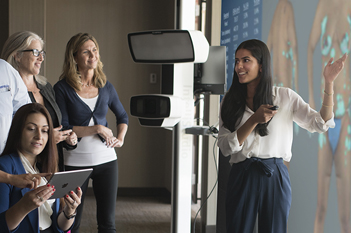

REVOLUTIONIZE
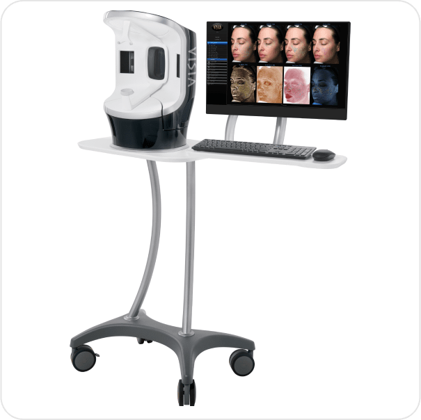
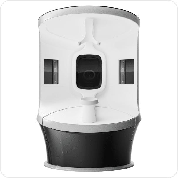
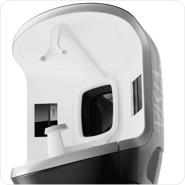
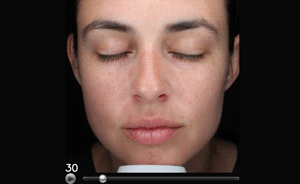
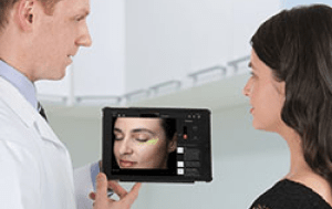
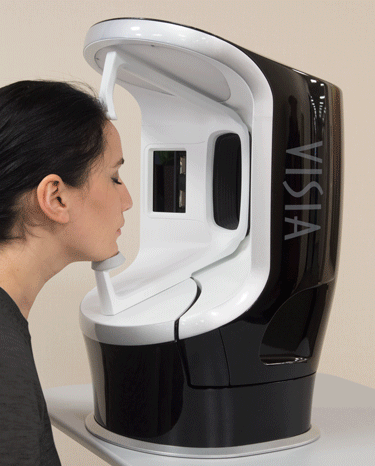
VISIA's capture module rotates smoothly around the subject to easliy photograph left, right and frontal facial views.
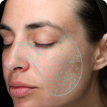
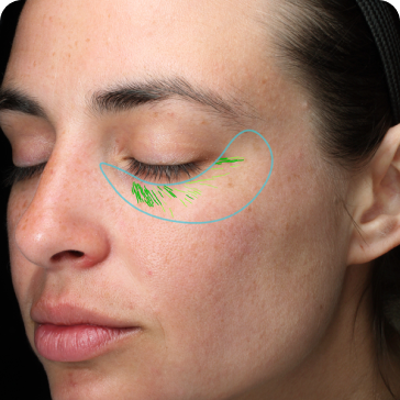
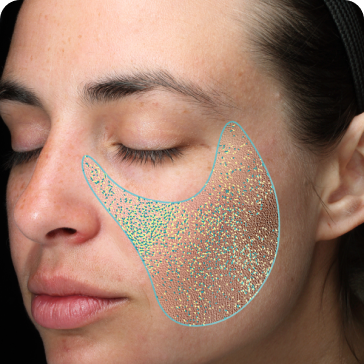
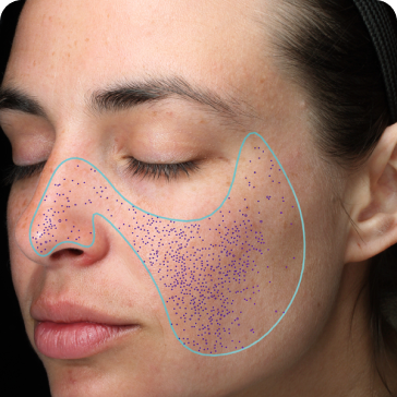
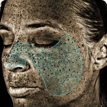
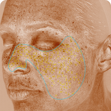
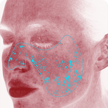
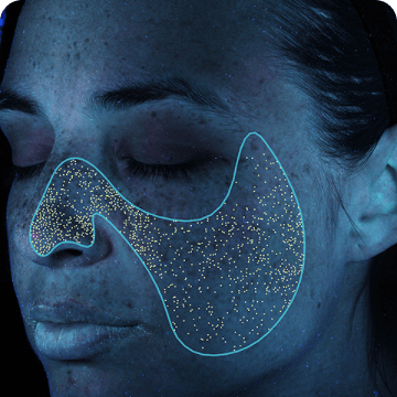
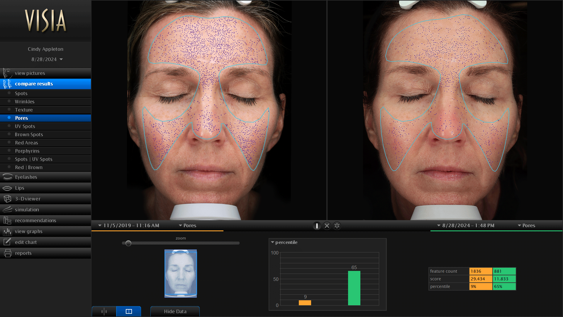
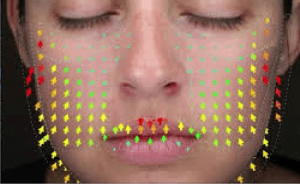
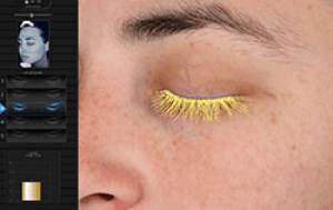
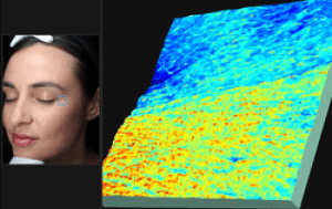
The multi-point positioning system and live image overlay make it easy to capture perfectly registered images to document progress over time.
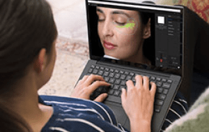
Simplify the imaging process while providing greater comfort for the client.